Research
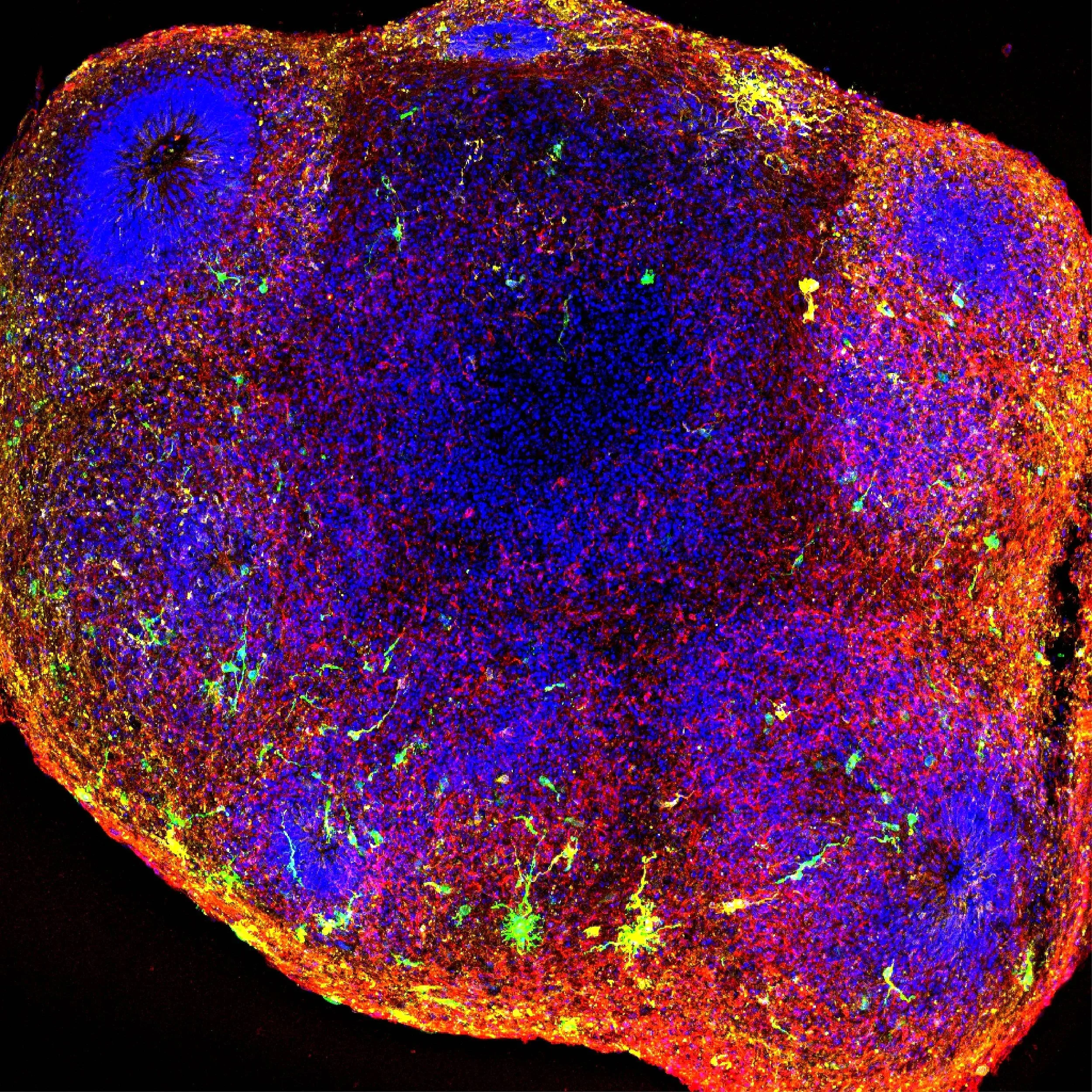
Tumor cells (green) engrafted into cortical organoids and stained for GFAP (yellow) and MAP2 (red).
HOW DO CANCER CELLS COMMUNICATE WITH NEURAL CELLS?
I study high-grade gliomas (HGGs), which are the most common brain tumors in adults. My particular focus is on glioblastoma, which is a highly aggressive and universally fatal brain tumor. Despite surgical removal, chemotherapy, and radiation therapy, almost all patients with glioblastoma will ultimately suffer from a relapse. One reason underlying a lack of curative options is that no two glioblastomas are alike, and even within a single tumor, malignant cells can occupy diverse cellular states and transition between them in response to their microenvironment. One feature that makes glioblastoma particularly aggressive is its ability to diffusively invade normal brain tissue in a tentacle-like manner. Within normal tissue, glioblastoma cells integrate into neural circuits and respond to neuronal activity as well as paracrine cues with an increase in proliferation and invasion. These modern discoveries echo histopathological findings from nearly a century ago when glioma cells were shown to cluster around neuronal cell bodies. Thus, glioblastoma tumors are not just masses that grow in the brain, but their biology is fundamentally intertwined with the neural microenvironment that surrounds them.
Another factor that makes glioblastoma difficult to treat is that these tumors are notorious shape-shifters that can morph into distinct cellular states to evade therapies. Intriguingly, many of these “states” reflect the same cell types, gene expression signatures, and pathways that emerge during normal brain development. For example, one of the major cell types within glioblastoma are radial glia—the neural progenitors that give rise to neurons and glial cells during development. These progenitors are also uniquely expanded in the human cerebral cortex, which is the site of ~90% of all adult gliomas. Thus, despite being an adult tumor, glioblastomas co-opt neurodevelopmental programs due to a pool of neural progenitors that remain dormant until later in life or a potential de-differentiation of cells in response to gene x environment interactions.
Together, these observations raise a central question: how does the neural microenvironment shape glioblastoma cellular identities, function, and therapeutic response? To address this, my goal is to develop clinically relevant and fully human model systems that recapitulate neural-tumor interactions in three dimensions. I accomplish this by collaborating closely with Emory’s Brain Organoid Hub, the Emory Department of Neurosurgery, and the newly-established Emory Winship Brain Tumor Center to obtain surgical resections of glioblastoma tissue directly from the operating room. I then engraft these patient-derived tumor cells into iPSC-derived, region-specific brain organoids and assembloids.
As many aspects of the human brain are not faithfully captured by traditional models, I believe these three-dimensional systems provide a novel and powerful platform to study patient-derived tumors within the uniquely human neural context in which they arise.
dd

Whole-mount imaging of neural-tumor connectivity in a human cortical organoid. Glioblastoma starter cells are in yellow, rabies-traced neurons in green, and nuclei in cyan.
MAIN QUESTIONS
1. Which neural circuits are most vulnerable to tumors?
Are these vulnerabilities random or pre-determined?
2. Do distinct neural circuits drive distinct tumor behaviors?
Can neural context reshape tumor cell identity?
3. How does manipulating the neural niche impact tumors?
How do injury, inflammation, and glial responses reshape tumor behavior?
4. Can neural-tumor interactions be therapeutically targeted?
Can we uncouple tumors from the neural circuits that sustain them?