Approaches
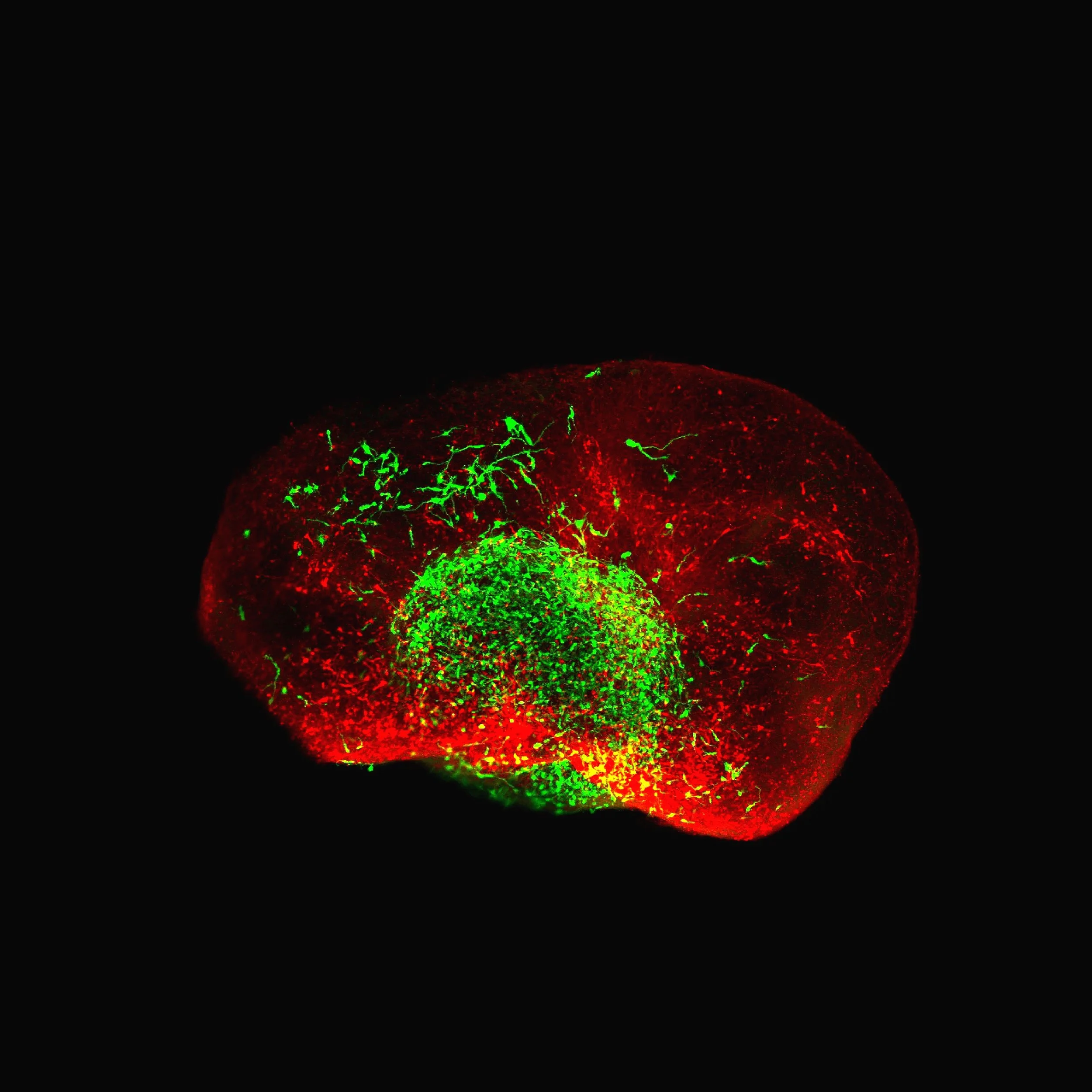
Whole glioblastoma patient-derived organoid (green) fused with a iPSC-derived cortical organoid (red) to form an assembloid-like system.
Human stem cell-derived 3D models & patient tumor tissues
My expertise is in reconstructing human neural environments using organoids & assembloids.
In this live-cell video, we can visualize engrafted patient-derived glioblastoma cells (green) interacting with corticospinal neurons (red) in an assembloid formed by fusing human cortical organoids with human spinal cord organoids.

I use monosynaptic tracers to map and manipulate neural-tumor circuits.
Shown in this whole-mount organoid stain are tumor cells expressing a receptor that allows selective infection with a glycoprotein-deleted rabies virus. Starter tumor cells are in red and green, while green-only cells identify their presynaptic partners.
Monosynaptic tracing techniques
I can visualize neural-tumor interactions across scales—from synapses to intact 3D tissues.
This greyscale video is of a tissue-cleared tumor-engrafted organoid imaged by confocal microscopy. We routinely pair these imaging approaches with optogenetic and chemogenetic perturbations to assess the functionality of neural-tumor circuits.
Functional imaging assays
Single-cell omics technologies & bioinformatics approaches
I leverage single-cell technologies to resolve how tumor and host cells adapt to the microenvironment.
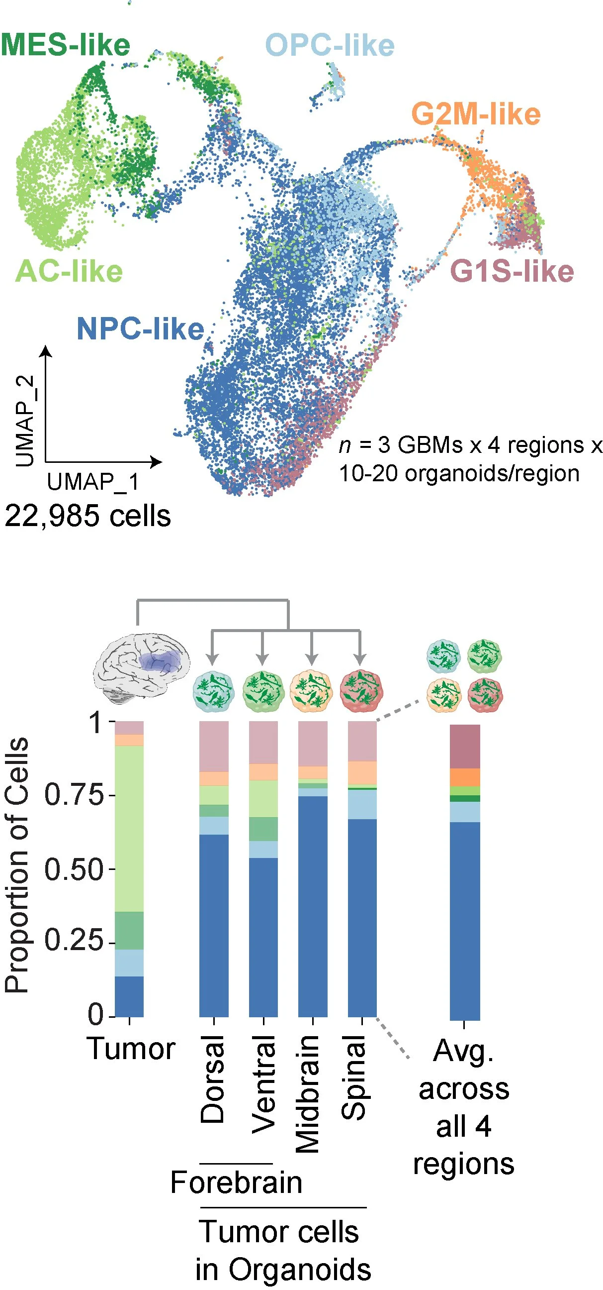
Described in this image is the inferred cellular makeup of resected glioblastomas and their cell state transitions after engraftment into brain organoids. We integrate such single-cell RNA sequencing data with bioinformatic pipelines of cell-cell communication as well as barcode-based lineage tracing to identify signaling pathways and track cell fate decisions.

Primary cell cultures & molecular biology
I validate our 3D model using 2D cell cultures, protein biochemistry, and molecular biology.
Shown are neurons immunopanned from human tissue and immunostained for TUJ1 (green) and MAP2 (red). We have extensive experience with such 2D cultures of neurons, astrocytes, and microglia isolated from human and rodent tissues, as well as surgically resected gliomas. We use these approaches along with immunoblotting and qPCR to validate our work in 3D models.
Postmortem human tissues, non-human primates & rodent models
I use complementary approaches spanning species & in vivo systems to confirm my findings.
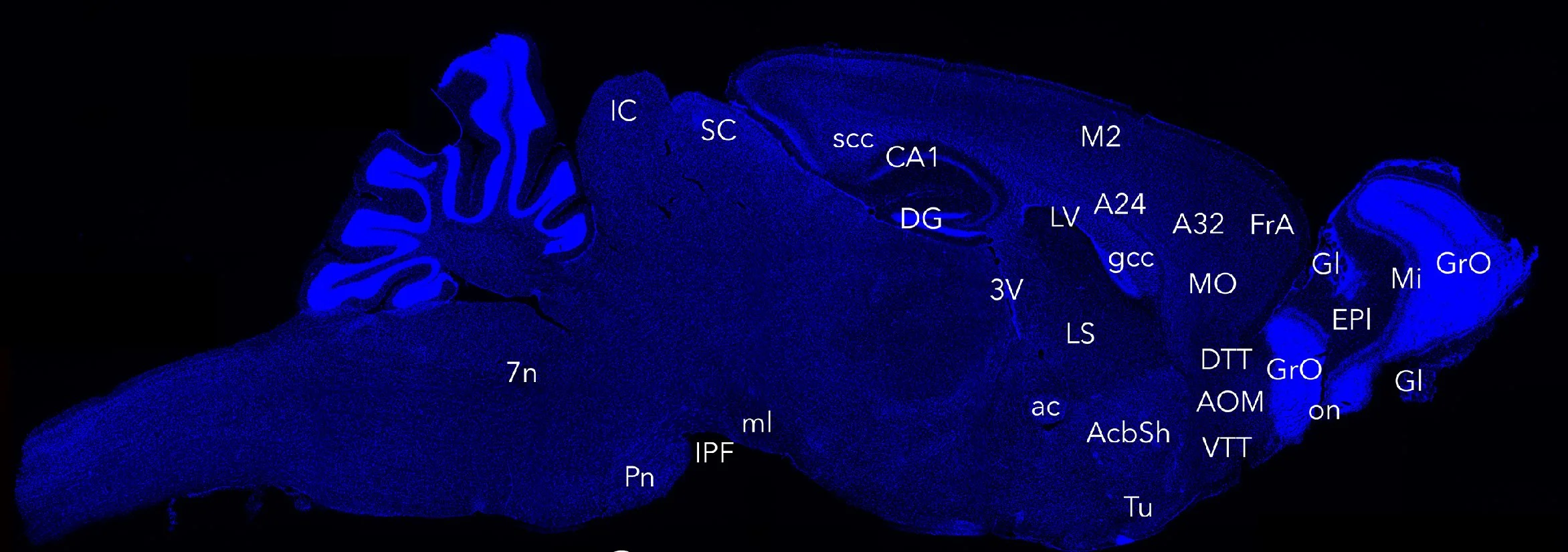
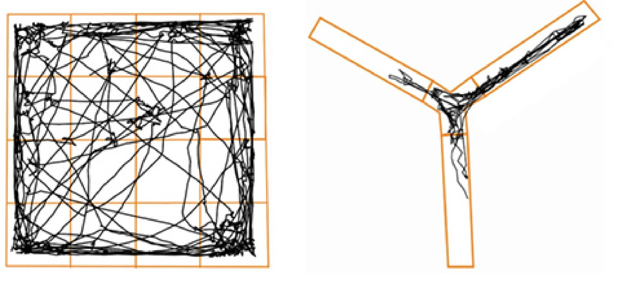
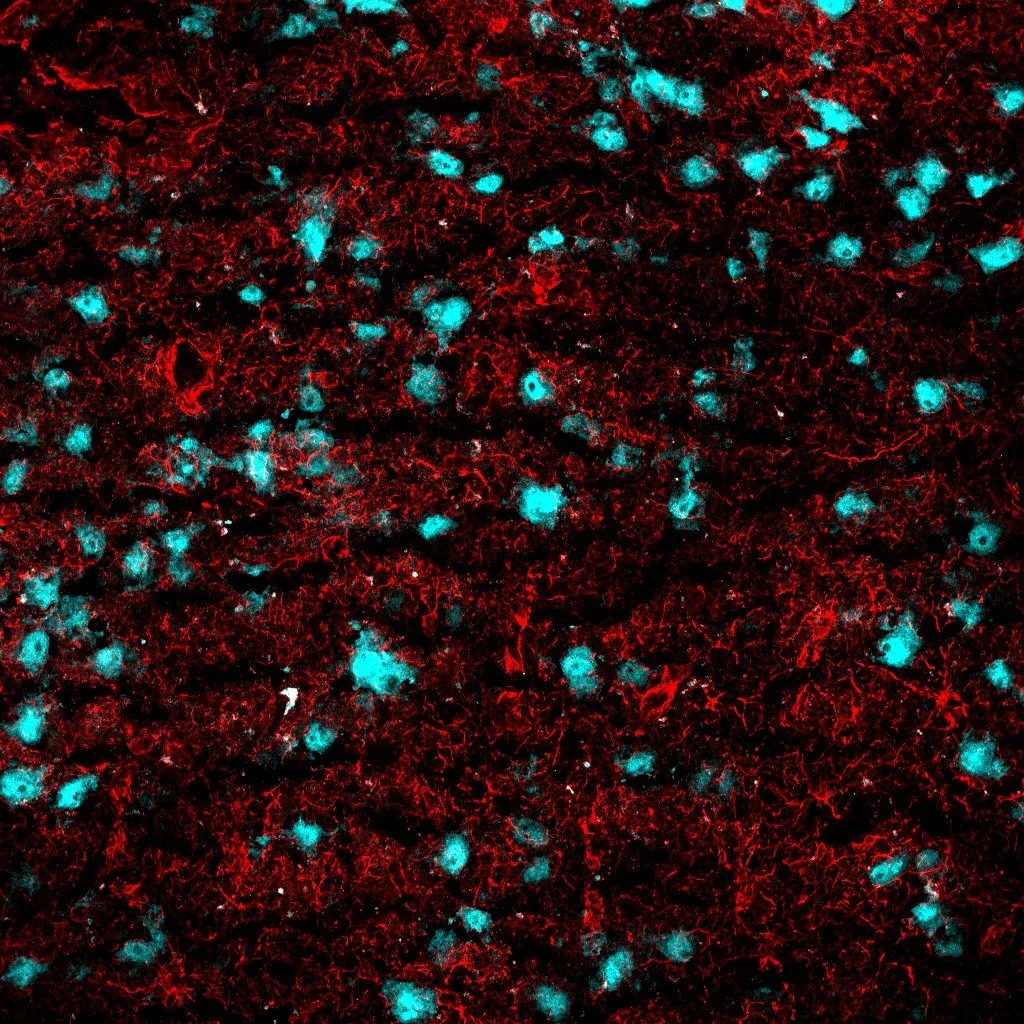
Shown here are a sagittal 20-month-old mouse brain section (top row), representative open-field and Y-maze behavioral track plots (middle row), & postmortem macaque brain tissue stained for NeuN (cyan) and GFAP (red). Our experience also includes stereotactic mouse brain surgery, analysis of brain-bank derived biospecimens, and behavioral assays of olfaction, cognition, and motor function.